A) Capitulum
B) Trochlea
C) Radial head and neck
D) Olecranon process
F) None of the above
Correct Answer

verified
Correct Answer
verified
Multiple Choice
Which of the following methods is used to demonstrate the carpal canal?
A) Stecher (PA axial)
B) Norgaard (AP oblique)
C) Lawrence (inferosuperior axial)
D) Gaynor-Hart (tangential)
F) A) and D)
Correct Answer

verified
Correct Answer
verified
Multiple Choice
How many phalanges are there in the hand?
A) 14
B) 27
C) 30
D) 32
F) B) and D)
Correct Answer

verified
Correct Answer
verified
Multiple Choice
The only saddle joint in the human body is the _____ joint.
A) radioulnar
B) radiocarpal
C) first digit,carpometacarpal
D) fifth digit,carpometacarpal
F) None of the above
Correct Answer

verified
Correct Answer
verified
Multiple Choice
The anatomy labeled as letter D in the figure above is the _____ joint. 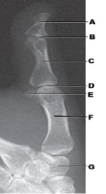
A) proximal IP
B) distal IP
C) metacarpophalangeal
D) carpometacarpal
F) All of the above
Correct Answer

verified
Correct Answer
verified
Multiple Choice
Which two bones comprise the forearm? (Select all that apply.)
A) Ulna
B) Fibula
C) Radius
D) Humerus
F) C) and D)
Correct Answer

verified
Correct Answer
verified
Multiple Choice
The position recommended to increase patient comfort when performing an AP projection of the humerus is:
A) prone.
B) recumbent.
C) supine.
D) upright.
F) B) and D)
Correct Answer

verified
Correct Answer
verified
Multiple Choice
Which two of the following should be demonstrated on the AP projection of the humerus? (Select all that apply.)
A) Wrist joint
B) Shoulder joint
C) Elbow joint
D) Entire clavicle
F) None of the above
Correct Answer

verified
Correct Answer
verified
Multiple Choice
What projection of the third digit is demonstrated in the figure above? 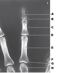
A) PA
B) PA oblique
C) Mediolateral
D) Lateromedial
F) A) and B)
Correct Answer

verified
Correct Answer
verified
Multiple Choice
The PA axial projection of the wrist (Stecher method) clearly demonstrates the:
A) lunate.
B) capitate.
C) scaphoid.
D) distal row of carpal bones.
F) A) and C)
Correct Answer

verified
Correct Answer
verified
Multiple Choice
What anatomy is labeled as letter D in the image below? 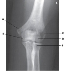
A) Lateral epicondyle of the humerus
B) Medial epicondyle of the humerus
C) Capitulum
D) Trochlea
F) All of the above
Correct Answer

verified
Correct Answer
verified
Multiple Choice
The carpal bones articulate with the:
A) radius only.
B) ulna only.
C) phalanges only.
D) radius,ulna,and phalanges.
F) All of the above
Correct Answer

verified
Correct Answer
verified
Multiple Choice
What bone is shown in this figure? 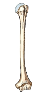
A) Radius
B) Tibia
C) Ulna
D) Humerus
F) C) and D)
Correct Answer

verified
Correct Answer
verified
Multiple Choice
Which of the following central-ray angles is used for the lateral projection of the wrist?
A) 0 degrees
B) 5 degrees
C) 7 degrees
D) 0 to 5 degrees
F) A) and C)
Correct Answer

verified
Correct Answer
verified
Multiple Choice
The general patient position most commonly used to perform a radiograph of a finger (digit) is:
A) AP.
B) PA.
C) sitting at the end of the table.
D) standing at the end of the table.
F) None of the above
Correct Answer

verified
Correct Answer
verified
Multiple Choice
Which of the following projections corrects foreshortening of the scaphoid?
A) PA
B) PA oblique in lateral rotation
C) PA in radial deviation
D) PA in ulnar deviation
F) A) and B)
Correct Answer

verified
Correct Answer
verified
Multiple Choice
To demonstrate the coronoid process in the axiolateral projection of the elbow (Coyle method) ,the elbow is flexed _____ degrees.
A) 45
B) 80
C) 90
D) 75
F) A) and D)
Correct Answer

verified
Correct Answer
verified
Multiple Choice
How many degrees is the central ray angled for the AP forearm?
A) 0
B) 5
C) 7
D) 3
F) All of the above
Correct Answer

verified
Correct Answer
verified
Multiple Choice
What projection is depicted in the image below? 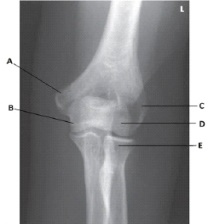
A) AP
B) AP oblique in medial rotation
C) AP oblique in lateral rotation
D) Mediolateral
F) A) and D)
Correct Answer

verified
Correct Answer
verified
Multiple Choice
What anatomy is labeled as letter A in the image below? 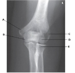
A) Lateral epicondyle of the humerus
B) Medial epicondyle of the humerus
C) Coronoid process of the ulna
D) Trochlea
F) B) and C)
Correct Answer

verified
Correct Answer
verified
Showing 41 - 60 of 136
Related Exams